Control iPSC line – iMA-1L
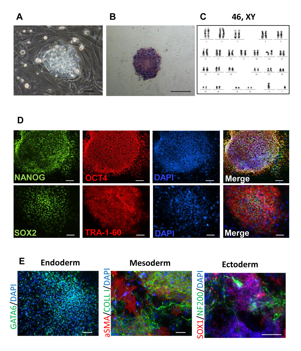
The induced pluripotent stem cell (iPSC) line iMA-1L was generated from human embryonic dermal fibroblasts using episomal vectors expressing pluripotency factors. The episomes were delivered into the cells by nucleofection (NHDF Nucleofector Kit, Lonza). iMA-1L intensively grew in the pluripotent stem cell-like colonies, had dense intercellular contacts, large nuclear-cytoplasmic ratio and expressed endogenous alkaline phosphatase. Immunofluorescent analysis for pluripotency markers showed the expression of OCT4, SOX2, NANOG, and TRA-1-60. iMA-1L has a normal karyotype 46: XY at both early (6) and late (22-24) passages. Immunofluorescent staining showed that iMA-1L can be differentiated into the cells of three germlines. The characteristics of this line was published in Supplementary Information in (Grigor’eva et al., 2020 https://doi.org/10.1007/s10616-020-00406-7).