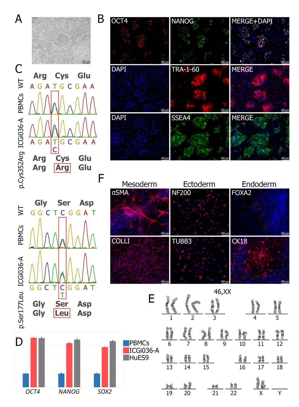
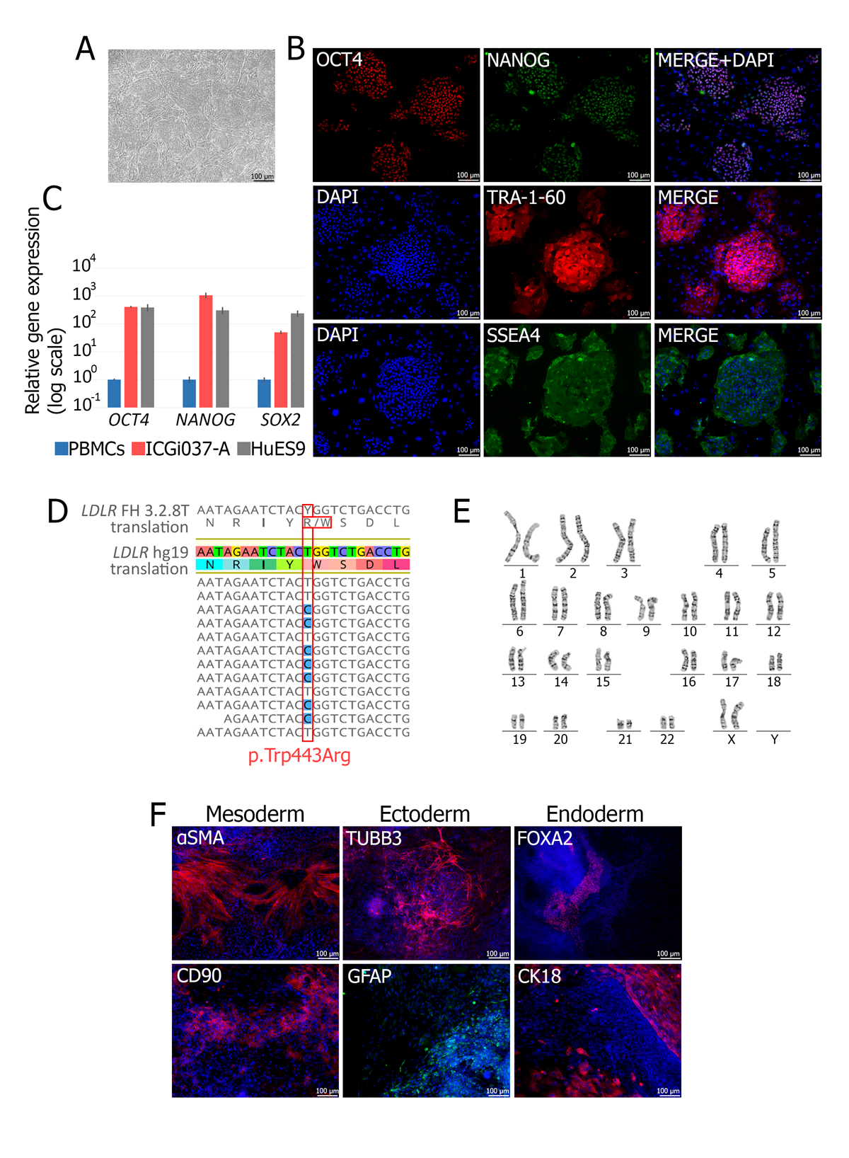
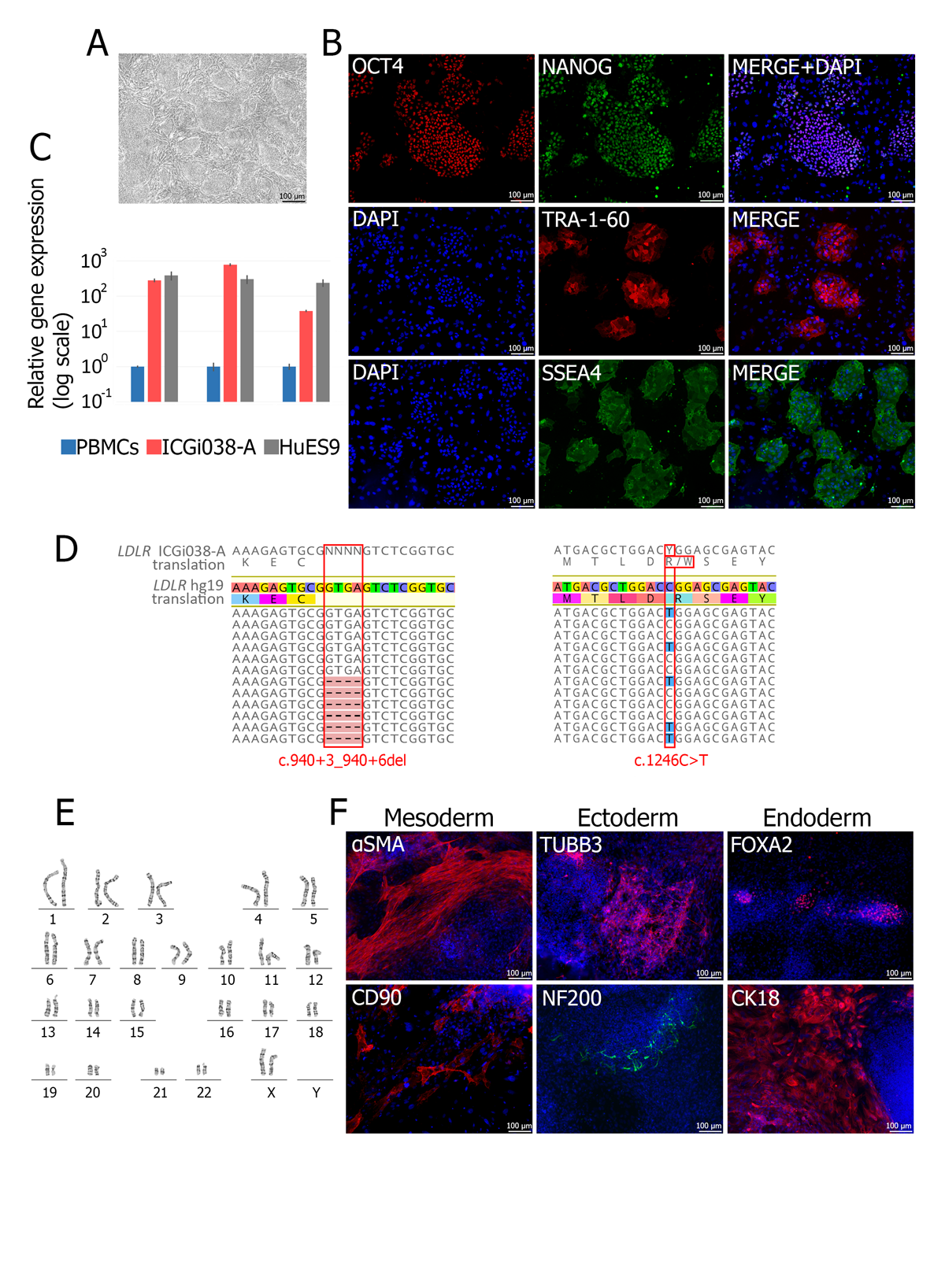
Familial hypercholesterolemia (FH) is a monogenic disease, leading to atherosclerosis due to a high level of low-density lipoprotein cholesterol. Most cases of the disease are based on pathological variants in the LDLR gene. Available treatments are effective not for all LDLR mutations. The development of cellular models for FH is an important direction for creating new approaches to atherosclerosis treatment.

This study was supported by the Russian Science Foundation (grant № 21-15-00065).

Diseased IPSC Lines-Familial Hypercholesterolemia_1
Characterization of the ICGi036-A iPSC line. A. Morphology of the iPSC colonies. B. Immunofluorescent staining for pluripotency markers OCT4, NANOG,TRA-1-60, SSEA4. C. Mutation analysis. Sanger sequencing of exons 7 and 4 of LDLR in the parental PBMCs and ICGi036-A iPSC line. Nucleotide substitutions areshown with red rectangles. D. Analysis of pluripotency genes (OCT4, NANOG, SOX2) expression in the iPSC line relative to parental PBMCs by RT-qPCR. Humanembryonic stem cell line, HUES9, was used as a control. E. Karyotype of the iPSC line. F. Immunofluorescent staining for three germ layer markers, αSMA andcollagen type I (mesoderm), NF200 and TUBB3 (ectoderm), CK18 and FOXA2/HNF3β (endoderm), after spontaneous in vitro differentiation in embryoid bodies.
Diseased IPSC Lines-Familial Hypercholesterolemia
Characterization of the ICGi037-A iPSC line. A. Morphology of the iPSC colonies. B. Immunofluorescent staining for pluripotency markers OCT4, NANOG,TRA-1-60, SSEA4. C. Analysis of pluripotency genes (OCT4, NANOG, SOX2) expression in the iPSC line relative to parental PBMCs by RT-qPCR. Human embryonicstem cell line, HUES9, was used as a control. D. Mutation analysis. Illumina HiSeq1500 sequencing of LDLR exon 9 in the ICGi037-A iPSC line. Nucleotide substitutionsare shown with red rectangles. E. Karyotype of the iPSC line. F. Immunofluorescent staining for three germ layer markers, αSMA and CD90 (mesoderm), TUBB3 and GFAP (ectoderm), CK18 and FOXA2/HNF3β (endoderm), after spontaneous in vitro differentiation in embryoid bodies.
IPSC-Diseased IPSC Lines-Familial Hypercholesterolemia
Characterization of the ICGi038-A iPSC line. A. Morphology of the iPSC colonies. B. Immunofluorescent staining for pluripotency markers OCT4, NANOG,TRA-1-60, SSEA4. C. Analysis of pluripotency genes (OCT4, NANOG, SOX2) expression in the iPSC line relative to parental PBMCs by RT-qPCR. Human embryonicstem cell line, HUES9, was used as a control. D. Mutation analysis. Illumina HiSeq1500 sequencing of LDLR in the ICGi038-A iPSC line. Nucleotide substitutions areshown with red rectangles. E. Karyotype of the iPSC line. F. Immunofluorescent staining for three germ layer markers, αSMA and CD90 (mesoderm), TUBB3 andNF200 (ectoderm), FOXA2/HNF3β and CK18 (endoderm), after spontaneous in vitro differentiation in embryoid bodies.